![]()
Created by
Paul A. Gulig, Ph.D.
Department of Molecular Genetics and Microbiology
with assistance from
David Brumbaugh
Here is a micrograph of the results of the Gram stain viewed at 1000X (oil immersion). Without oil immersion to limit light diffraction, you cannot tell much about the morphology or even color of the bacterial cells. Both colony types 1 and 2 are identical by the Gram stain, so one picture is shown for both.
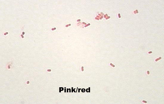 They are gram-negative rods.