Key to Lab Images
Here is a set of images
from the four cases.
The first four images are
what the blood agar
plates from the four cases looked like:
 Case
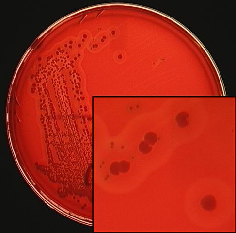
1 contains large somewhat beta-hemolytic colonies and small
alpha-hemolytic colonies. There are some larger colonies that might
have appeared in the heavy regions of the streaking pattern, but
probably
not in the isolated area. The enlarged inset area shows the different
colony types in more detail.
Case
1 contains large somewhat beta-hemolytic colonies and small
alpha-hemolytic colonies. There are some larger colonies that might
have appeared in the heavy regions of the streaking pattern, but
probably
not in the isolated area. The enlarged inset area shows the different
colony types in more detail.
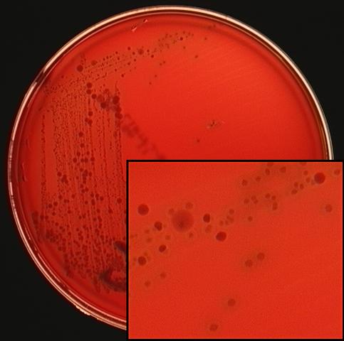 Case
2 contains small, white
non-hemolytic colonies and small
alpha-hemolytic
colonies. The same large colonies from Case 1 are present in the heavy
streak
area. The enlarged inset area shows the different colony types in more
detail.
Case
2 contains small, white
non-hemolytic colonies and small
alpha-hemolytic
colonies. The same large colonies from Case 1 are present in the heavy
streak
area. The enlarged inset area shows the different colony types in more
detail.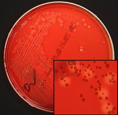 Case
3 contains small heavily beta-hemolytic colonies and small, white
non-hemolytic colonies. The same large colonies from Case 1 are present
in the
heavy streak area. The enlarged inset area shows the different colony
types in
more detail.
Case
3 contains small heavily beta-hemolytic colonies and small, white
non-hemolytic colonies. The same large colonies from Case 1 are present
in the
heavy streak area. The enlarged inset area shows the different colony
types in
more detail.
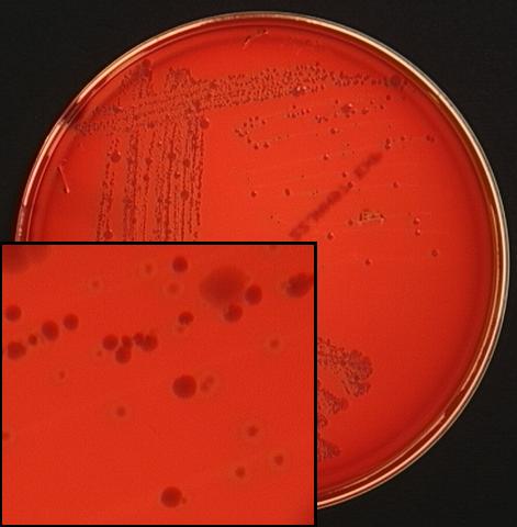 Case
4 contains small, white
non-hemolytic colonies and small
alpha-hemolytic
colonies. The same large colonies from Case 1 are present in the heavy
streak
area. The enlarged inset area shows the different colony types in more
detail.
Case
4 contains small, white
non-hemolytic colonies and small
alpha-hemolytic
colonies. The same large colonies from Case 1 are present in the heavy
streak
area. The enlarged inset area shows the different colony types in more
detail.
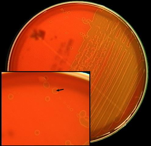 Alpha
hemolysis is shown more clearly
in this blood agar plate of
Streptococcus
pneumoniae in pure
culture. The arrow in the inset
shows that there is a very small colony surrounded by an olive green
discoloration and a thin, lighter halo of hemolysis.
Alpha
hemolysis is shown more clearly
in this blood agar plate of
Streptococcus
pneumoniae in pure
culture. The arrow in the inset
shows that there is a very small colony surrounded by an olive green
discoloration and a thin, lighter halo of hemolysis.
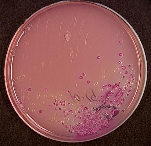
All of the cases looked about the same on MacConkey
Agar - about an equal mix of Lac+ and Lac-colonies, but very few in
number
compared with the colonies seen on the blood agar plates. The colonies
observed
from the MacConkey agar
were "lost" in the
heavy portion of the blood agar. Why were they clearly visible on the
MacConkey agar?
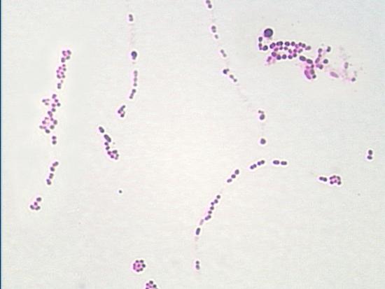
The following images are representative Gram stains of the morphologies from the unknowns.
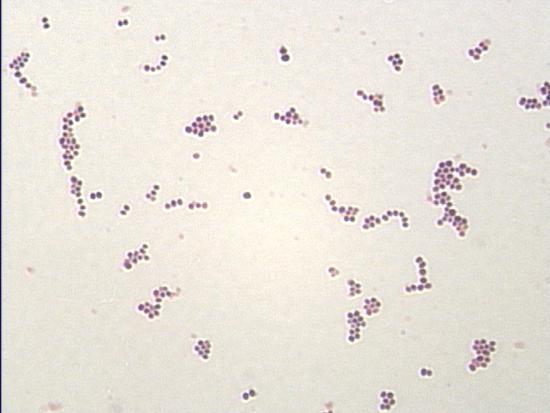 Staphylococcus
aureus gram-positive cocci usually in bunches, but not always.
Staphylococcus
aureus gram-positive cocci usually in bunches, but not always.
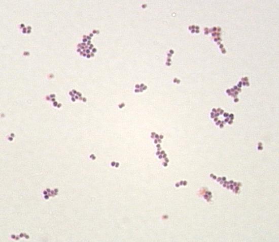 Staphylococcus
epidermidis gram-positive cocci usually in bunches, but not always.
Note that you cannot distinguish the staphylococci by the Gram stain.
Staphylococcus
epidermidis gram-positive cocci usually in bunches, but not always.
Note that you cannot distinguish the staphylococci by the Gram stain.
 Streptococcus
pyogenes
(Group A strep) gram-positive
cocci usually in chains, but not always.
Streptococcus
pyogenes
(Group A strep) gram-positive
cocci usually in chains, but not always.
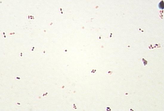 Streptococcus
pneumoniae
gram-positive diplococci.
Notice the characteristic elongated shape of the diplococci.
Streptococcus
pneumoniae
gram-positive diplococci.
Notice the characteristic elongated shape of the diplococci.
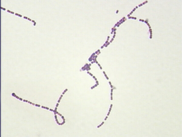 Viridans
streptococci
gram-positive cocci usually in
chains, but not always. Notice that these cannot be differentiated from
Streptococcus pyogenes
by the Gram stain.
Viridans
streptococci
gram-positive cocci usually in
chains, but not always. Notice that these cannot be differentiated from
Streptococcus pyogenes
by the Gram stain.
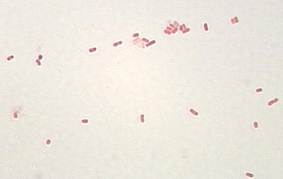 All
of the gram-negative rods look the
same.
All
of the gram-negative rods look the
same.
The following images are what some of the differential tests should
look like:
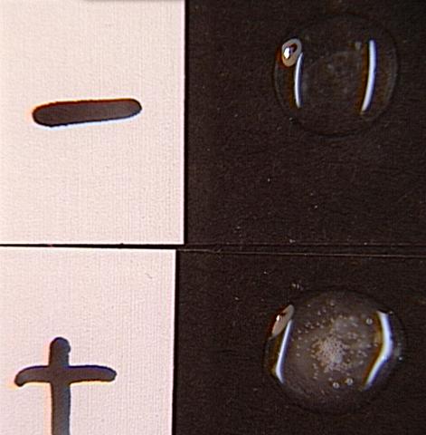 The
catalase test Note the bubbles in
the lower positive test.
The
catalase test Note the bubbles in
the lower positive test.
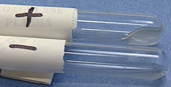 The
coagulase test Note that the upper
positive reaction has gelled,
while the
lower negative reaction is liquid.
The
coagulase test Note that the upper
positive reaction has gelled,
while the
lower negative reaction is liquid.
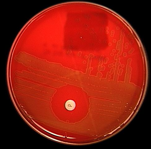
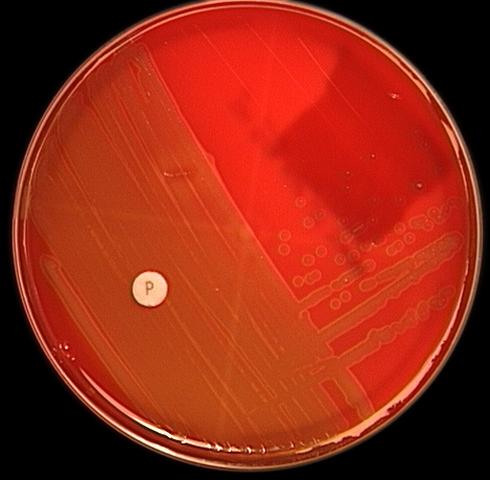 The optochin (P
disk) test for identifying Streptococcus pneumoniae. Not the zone of
inhibition of growth for Streptococcus pneumoniae and the lack of
inhibition for viridans streptococci.
The optochin (P
disk) test for identifying Streptococcus pneumoniae. Not the zone of
inhibition of growth for Streptococcus pneumoniae and the lack of
inhibition for viridans streptococci.
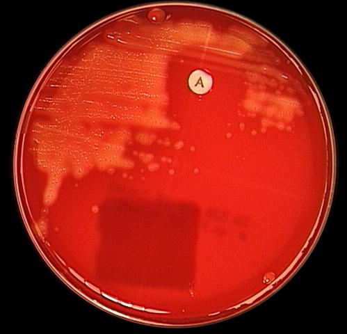 The
bacitracin (A disk) test for
identifying Streptococcus pyogenes. Note the zone of inhibition of
growth for Streptococcus pyogenes.
The
bacitracin (A disk) test for
identifying Streptococcus pyogenes. Note the zone of inhibition of
growth for Streptococcus pyogenes.
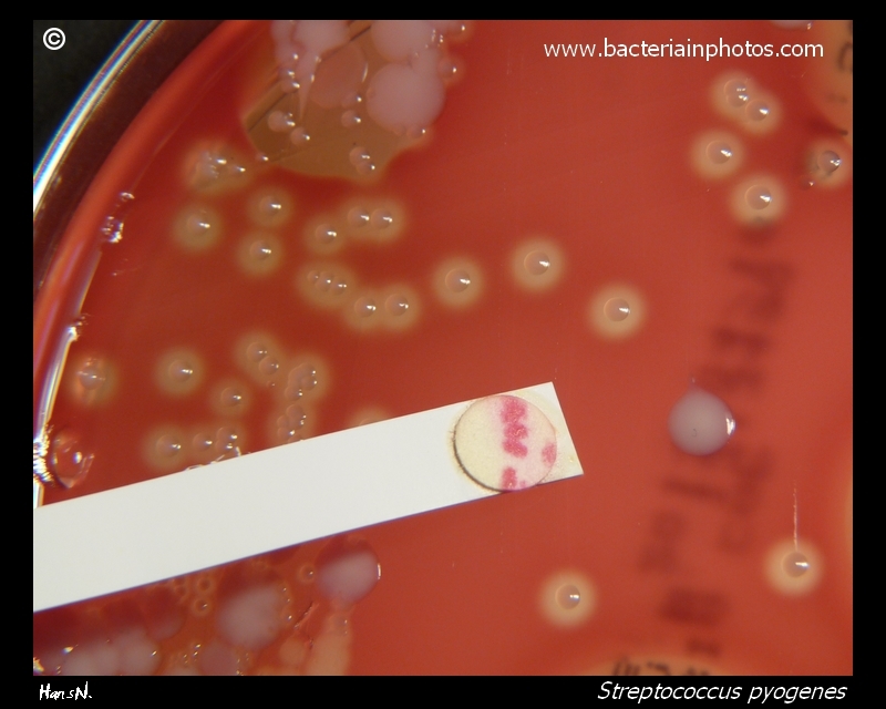
The
Binax Strep A point-of-care test. The lower line is the test line
that has the anti-Group A carbohydrate detection antobody. A red
line here shows that the gold particles with anti-Group A antibodies
had bound to Group A antigen in the sample and were captured by the
test antibody. The upper band is the control line that has
anti-IgG/IgM antibodies. These antibodies will bind the gold
particles with anti-Group A antibodies regardless of if there is Group
A antigen inthe sample or not. If this line is not red, the test
is invalid.

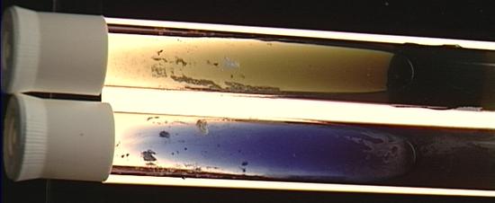 The
citrate test is used in this lab exercise to differentiate E. coli from
Klebsiella
pneumoniae. In real life
there is much more involved
than this simple test. The upper tube is a negative reaction with the
original green color remaining, while the lower tube is a positive
reaction with
the green having changed to blue.
The
citrate test is used in this lab exercise to differentiate E. coli from
Klebsiella
pneumoniae. In real life
there is much more involved
than this simple test. The upper tube is a negative reaction with the
original green color remaining, while the lower tube is a positive
reaction with
the green having changed to blue.
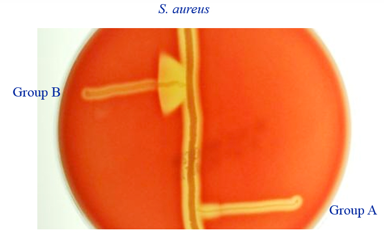
CAMP test. The CAMP test is used to differentiate beta-hemolytic streptococci. Group B strep (S. agalactiae) is CAMP-positive, but Group A strep (S. pyogenes) is CAMP-negative.Staphylococcus aureus is streaked on a blood agar plate, and the unknown beta-hemolytic strep is cross-streaked across the staph. An arrowhead-shaped zone of heightened hemolysis at the junction is a positive result.