![]()
Created by
Paul A. Gulig, Ph.D.
Department of Molecular Genetics and Microbiology
with assistance from
David Brumbaugh
Here again is an image of the blood agar plate from case 2. Now let's work up colony type #2, which is to the lower left of the #2.
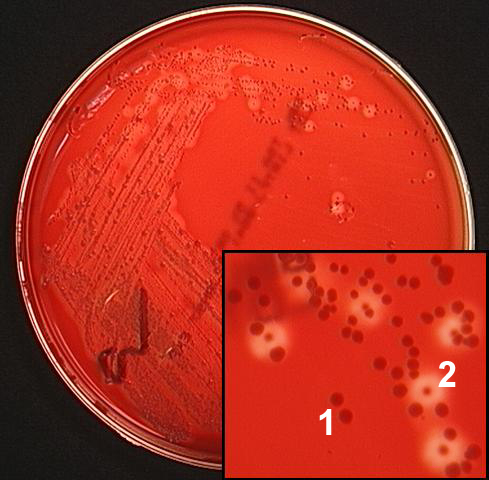 How would you describe colony #2?
A. Beta-hemolytic and large.
B. Alpha hemolytic and small.
C. Gamma-hemolytic and large.
D. Beta-hemolytic and small.
What will you do next with colony #2?
A. Test for antibiotic resistance.
B. Perform a Gram stain.
C. Do a PCR.
D. All of the above.